Mr Andrew Mahon | Common Wrist Conditions
16742
page-template,page-template-full_width,page-template-full_width-php,page,page-id-16742,page-child,parent-pageid-16720,ajax_fade,page_not_loaded,,qode_grid_1300,footer_responsive_adv,qode-theme-ver-9.1.3,wpb-js-composer js-comp-ver-4.11.2.1,vc_responsive
 

Common Wrist Conditions

The information outlined below on common conditions and treatments is provided as a guide only and it is not intended to be comprehensive.


Discussion with Mr Mahon is important to answer any questions that you may have. For information about any additional conditions not featured within the site, please contact us for more information.

COMMON CONDITIONS OF THE WRIST
CARPAL TUNNEL SYNDROME

What is it?


Carpal tunnel syndrome (CTS) is a condition where the median nerve is compressed where it passes through a short tunnel at the wrist. The tunnel contains the tendons that bend the fingers and thumb as well as the nerve (see diagram). CTS commonly affects women in middle age but can occur at any age in either sex. CTS can occur with pregnancy, diabetes, thyroid problems, rheumatoid arthritis and other less common conditions, but most sufferers have none of these. CTS may be associated with swelling in the tunnel which may be caused by inflammation of the tendons, a fracture of the wrist, wrist arthritis and other less common conditions. In most cases, the cause is not identifiable.


What are the symptoms?


The main symptom is altered feeling in the hand, affecting the thumb index, middle and ring fingers; it is unusual for the little finger to be involved. Many people describe the altered feeling as tingling. Tingling is often worse at night or first thing in the morning. It may be provoked by activities that involve gripping an object, for example a mobile telephone or newspaper, especially if the hand is elevated. In the early stages the symptoms of tingling intermittent and sensation will return to normal. If the condition worsens, the altered feeling may become continuous, with numbness in the fingers and thumb together with weakness and wasting of the muscles at the base of the thumb. Sufferers often described a feeling of clumsiness and drop objects easily. CTS may be associated with pain in the wrist and forearm.


In some cases, nerve conduction tests are needed to confirm the diagnosis. Blood tests and x-rays are sometimes required.


What is the treatment?


Non-surgical treatments include the use of splints, especially at night, and steroid injection into the carpal tunnel. CTS occurring in pregnancy often resolves after the baby is born.


Surgery is frequently required. The operation involves opening the roof of the tunnel to reduce the pressure on the nerve (see diagram: the roof of the carpal tunnel is called the transverse carpal ligament). The most common method involves an incision over the tunnel at the wrist, opening the roof under direct vision. In an alternative keyhole method (endoscopic release) the roof is opened with instruments inserted through one or two small incisions. The outcomes of the two techniques are similar and your surgeon can discuss the most appropriate method. The surgery may be performed under local anaesthesia, regional anaesthesia (injected at the shoulder to numb the entire arm) or general anaesthesia.


The outcome is usually a satisfactory resolution of the symptoms. Night pain and tingling usually disappear within a few days. In severe cases, improvement of constant numbness and muscle weakness may be slow or incomplete. It generally takes about three months to regain full strength and a fully comfortable scar, but the hand can be used for light activities from the day of surgery.

DISTAL RADIUS FRACTURES

The radius is the larger of the two bones of the forearm. The end toward the wrist is called the distal end. A fracture of the distal radius occurs when the area of the radius near the wrist breaks.


Distal radius fractures are very common. In fact, the radius is the most commonly broken bone in the arm. A distal radius fracture almost always occurs about 1 inch from the end of the bone. The break can occur in many different ways, however.


One of the most common distal radius fractures is a Colles fracture, in which the broken fragment of the radius tilts upward. This fracture was first described in 1814 by an Irish surgeon and anatomist, Abraham Colles — hence the name “Colles” fracture. A Colles fracture occurs when the broken end of the radius tilts upward.


Other ways the distal radius can break include:


Intra-articular fracture. A fracture that extends into the wrist joint. (“Articular” means “joint.”)
Extra-articular fracture. A fracture that does not extend into the joint is called an extra-articular fracture.
Open fracture. When a fractured bone breaks the skin, it is called an open fracture. These types of fractures require immediate medical attention because of the risk for infection.
Comminuted fracture. When a bone is broken into more than two pieces, it is called a comminuted fracture.


It is important to classify the type of fracture, because some fractures are more difficult to treat than others. Intra-articular fractures, open fractures, comminuted fractures, and displaced fractures (when the broken pieces of bone do not line up straight).are more difficult to treat, for example.


Sometimes, the other bone of the forearm (the ulna) is also broken. This is called a distal ulna fracture.


What is the cause ?


The most common cause of a distal radius fracture is a fall onto an outstretched arm.


Osteoporosis (a disorder in which bones become very fragile and more likely to break) can make a relatively minor fall result in a broken wrist. Many distal radius fractures in people older than 60 years of age are caused by a fall from a standing position.


A broken wrist can happen even in healthy bones, if the force of the trauma is severe enough. For example, a car accident or a fall off a bike may generate enough force to break a wrist.


Good bone health remains an important prevention option. Wrist guards may help to prevent some fractures, but they will not prevent them all.


What are the symptoms ?


A broken wrist usually causes immediate pain, tenderness, bruising, and swelling. In many cases, the wrist hangs in an odd or bent way (deformity).


What is the treatment ?


Treatment of broken bones follows one basic rule: the broken pieces must be put back into position and prevented from moving out of place until they are healed.


There are many treatment options for a distal radius fracture. The choice depends on many factors, such as the nature of the fracture, your age and activity level, and the surgeon’s personal preferences.


Nonsurgical Treatment


If the broken bone is in a good position, a plaster cast may be applied until the bone heals.


If the position (alignment) of your bone is out of place and likely to limit the future use of your arm, it may be necessary to re-align the broken bone fragments. “Reduction” is the technical term for this process in which the doctor moves the broken pieces into place. When a bone is straightened without having to open the skin (incision), it is called a closed reduction.


After the bone is properly aligned, a splint or cast may be placed on your arm. A splint is usually used for the first few days to allow for a small amount of normal swelling. A cast is usually added a few days to a week or so later, after the swelling goes down. The cast is changed 2 or 3 weeks later as the swelling goes down more, causing the cast to loosen.


Depending on the nature of the fracture, your doctor may closely monitor the healing by taking regular x-rays. . If the fracture was reduced or thought to be unstable, x-rays may be taken at weekly intervals for 3 weeks and then at 6 weeks. X-rays may be taken less often if the fracture was not reduced and thought to be stable.


The cast is removed about 6 weeks after the fracture happened. At that point, physical therapy is often started to help improve the motion and function of the injured wrist.


Surgical Treatment


Sometimes, the position of the bone is so much out of place that it cannot be corrected or kept corrected in a cast. This has the potential of interfering with the future functioning of your arm. In this case, surgery may be required.


Procedure. Surgery typically involves making an incision to directly access the broken bones to improve alignment (open reduction).


Depending on the fracture, there are a number of options for holding the bone in the correct position while it heals:


Cast
Metal pins (usually stainless steel or titanium)
Plate and screws
External fixator (a a stabilizing frame outside the body that holds the bones in the proper position so they can heal)
Any combination of these techniques


Open fractures. Surgery is required as soon as possible (within 8 hours after injury) in all open fractures. The exposed soft tissue and bone must be thoroughly cleaned (debrided) and antibiotics may be given to prevent infection. Either external or internal fixation methods will be used to hold the bones in place. If the soft tissues around the fracture are badly damaged, your doctor may apply a temporary external fixator. Internal fixation with plates or screws may be utilized at a second procedure several days later.


What is the outcome?
Because the kinds of distal radius fractures are so varied and the treatment options are so broad, recovery is different for each individual. Talk to Mr Mahon for specific information about your recovery program and return to daily activities.

NERVE DAMAGE

A nerve injury can occur when the wrist is cut, overstretched, crushed or burned. Symptoms include numbness and difficulty moving the affected area.


Treatment depends on the severity of the injury, and may include rest, immobilization, physical therapy, and in some cases, surgery.


Causes of hand & wrist nerve damage


Injury to nerves in the wrist is often caused by trauma, such as the area being cut, overstretched, crushed or burned, damaging the nerve or nerve ending.


Symptoms of hand & nerve damage


The nerves located in the wrist affect motor and sensory functions. If a nerve in the wrist is cut, severed, stretched, or otherwise damaged, it will not function properly (or at all)


Damage to motor nerves can cause weakness, twitching, and paralysis in the hand, wrist and arm. Sensory nerve damage symptoms include pain, numbness, tingling, increased sensitivity, burning, and problems positioning the hand correctly.


Similarly, when a nerve in the hand or wrist is pinched or compressed (such as in carpal tunnel syndrome), there can be pain and loss of feeling through the hand and fingers.


Treatment of hand & nerve damage


If the nerve is pinched or compressed, surgery may be able to release the nerve and restore functionality, depending on the severity of damage and how long the nerve was compressed.

SCAPHOID FRACTURES

A scaphoid (navicular) fracture is a break in one of the small bones of the wrist. This type of fracture occurs most often after a fall onto an outstretched hand. Symptoms of a scaphoid fracture typically include pain and tenderness in the area just below the base of the thumb. These symptoms may worsen when you try to pinch or grasp something.


Treatment for a scaphoid fracture can range from casting to surgery, depending on the fracture’s severity and location on the bone. Because portions of the scaphoid have a poor blood supply—and a fracture can further disrupt the flow of blood to the bone—complications with the healing process are common.


A scaphoid fracture is usually described by its location within the bone. Most commonly, the scaphoid breaks in its mid-portion, called the “waist.” Fractures can also occur at both the proximal and distal ends of the bone.


Scaphoid fractures are classified according to the severity of displacement–or how far the pieces of bone have moved out of their normal position:


Non-displaced fracture. In this type of fracture, the bone fragments line up correctly.
Displaced fracture. In this type of fracture, the bone fragments have moved out of their normal position. There may be gaps between the pieces of bone or fragments may overlap each other.


What is the cause ?


A scaphoid fracture usually occurs when you fall onto an outstretched hand, with your weight landing on your palm. The end of the larger forearm bone (the radius) may also break in this type of fall, depending on the position of the hand on landing.


The injury can also happen during sports activities or motor vehicle collisions.


Fractures of the scaphoid occur in people of all ages, including children.There are no specific risk factors or diseases that make you more likely to experience a scaphoid fracture. Some studies have shown that using wrist guards during high-energy activities like inline skating and snowboarding can help decrease your chance of breaking a bone around the wrist.


What are the symptoms ?


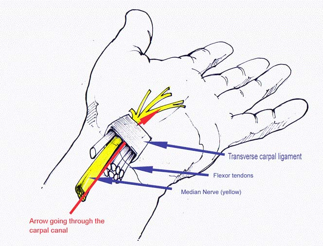
Scaphoid fractures usually cause pain and swelling in the anatomic snuffbox and on the thumb side of the wrist. The pain may be severe when you move your thumb or wrist, or when you try to pinch or grasp something. Unless your wrist is deformed, it might not be obvious that your scaphoid bone is broken. With some scaphoid fractures, the pain is not severe and may be mistaken for a wrist sprain.


Pain in your wrist that does not go away within a day of injury may be a sign of a fracture—so it is important to see a doctor if your pain persists. Prompt treatment of a scaphoid fracture will help avoid potential complications.


What is the treatmemt ?


The treatment your doctor recommends will depend on a number of factors, including:


The location of the break in the bone
Whether the bone fragments are displaced
How long ago your injury occurred


Nonsurgical Treatment


Fracture near the thumb. Scaphoid fractures that are closer to the thumb (distal pole) usually heal in a matter of weeks with proper protection and restricted activity. This part of the scaphoid bone has a good blood supply, which is necessary for healing. For this type of fracture, your doctor may place your forearm and hand in a cast or a splint. The cast or splint will usually be below the elbow and include your thumb. Healing time varies from patient to patient. Your doctor will monitor your healing with periodic x-rays or other imaging studies.


Fracture near the forearm. If the scaphoid is broken in the middle of the bone (waist) or closer to the forearm (proximal pole), healing can be more difficult. These areas of the scaphoid do not have a very good blood supply. If your doctor treats this type of fracture with a cast, the cast may include the thumb and extend above the elbow to help stabilize the fracture


Bone stimulator. In some cases, your doctor may recommend the use of a bone stimulator to assist in fracture healing. This small device delivers low-intensity ultrasonic or pulsed electromagnetic waves that stimulate healing.


Surgical Treatment


If your scaphoid is broken at the waist or proximal pole or if pieces of bone are displaced, your doctor may recommend surgery. The goal of surgery is to realign and stabilize the fracture, giving it a better chance to heal.


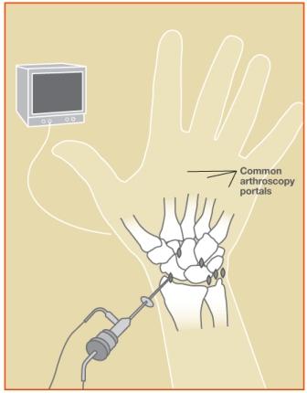
Reduction. During this procedure, your doctor will administer an anesthetic or anesthesia and manipulate the bone back into its proper position. In some cases, this is done using a limited incision and special guided instruments. In other cases, it is performed through an open incision with direct manipulation of the fracture. For some fractures, your doctor may use a tiny camera called an “arthroscope” to aid in the reduction.


Internal fixation. During this procedure, metal implants—including screws and/or wires—are used to hold the scaphoid in place until the bone is fully healed. The location and size of the surgical incision depends on what part of the scaphoid is broken. Sometimes, the screw or wire can be placed in bone fragments with a small incision. In other cases, a larger incision is needed to ensure that the fragments of the scaphoid line up properly. The incision may be made on either the front or the back of your wrist.


Bone graft. In some cases, a bone graft may be used with or without internal fixation. A bone graft is new bone that is placed around the broken bone. It can stimulate bone production and healing. The graft may be taken from your forearm bone in the same arm or from your hip.


What is the outcome


Whether your treatment is surgical or nonsurgical, you may be required to wear a cast or splint for up to 6 months or until your fracture has healed. Unlike most other fractures, scaphoid fractures tend to heal slowly. During this time, unless advanced activity is approved by your doctor, you should avoid the following activities:


Lifting, carrying, pushing, or pulling more than one pound of weight
Throwing with your injured arm
Participating in contact sports
Climbing ladders or trees
Participating in activities with a risk of falling onto your hand, such as inline skating or jumping on a trampoline
Using heavy or vibratory machinery
Smoking (which can delay or prevent fracture healing)


Some patients have wrist stiffness after scaphoid fractures. This is more common in patients who wear a cast for a long time or require more extensive surgery.


It is important to maintain full finger motion throughout your recovery period. Your doctor will provide an exercise program, and may refer you to a trained hand therapist who will help you regain as much range of motion and strength in your wrist as possible.


Despite hand therapy and a great deal of effort by the patient during home therapy, some patients may not recover the same range of motion and strength that they had before their injury.

TENDON / LIGAMENT REPAIR

If any of the tendons in your hand are damaged, surgery may be needed to repair them and help restore movement in the affected fingers or thumb.


What are tendons?


Tendons are tough cords of tissue that connect muscles to bones. When a group of muscles contract (tighten), the attached tendons will pull on certain bones, allowing you to make a wide range of movements.


There are two groups of tendons in the hand:


▪ extensor tendons – which run from the forearm, across the back of your hand to your fingers and thumb, allowing you to straighten your fingers and thumb


▪ flexor tendons – which run from your forearm, through your wrist and across the palm of your hand, allowing you to bend your fingers.


Surgery can often be carried out to repair damage to both these groups of tendons.


When hand tendon repair is needed Hand tendon repair is carried out when one or more tendons in your hand rupture or are cut, leading to loss of normal hand movements.


If your extensor tendons are damaged, you’ll be unable to straighten one or more fingers. If your flexor tendons are damaged, you’ll be unable to bend one or more fingers. Tendon damage can also cause pain and inflammation (swelling) in your hand.


In some cases, damage to the extensor tendons can be treated without the need for surgery, using a rigid support called a splint thatʼs worn around the hand.


Common causes of tendon injuries include:


• cuts – cuts across the back or palm of your hand can result in injury to your tendons


• sports injuries – extensor tendons can rupture when stubbing a finger, such as trying to catch a ball; flexor tendons can occasionally be pulled off the bone when grabbing an opponent’s jersey, such as in rugby; and the pulleys holding flexor tendons can rupture during activities that involve lots of strenuous gripping, such as rock climbing


• bites – animal and human bites can cause tendon damage, and a person may damage their hand tendon after punching another person in the teeth


• crushing injuries – jamming a finger in a door or crushing the hand in a car accident can divide or rupture a tendon


• rheumatoid arthritis – rheumatoid arthritis can cause tendons to become inflamed, which in severe cases can lead to tendons rupturing


Tendon repair surgery


Tendon repair may involve the surgeon making an incision in your wrist, hand or finger so they can locate the ends of the divided tendon and stitch them together. Extensor tendons are easier to reach, so repairing them is relatively straightforward. Depending on the type of injury, it may be possible to repair extensor tendons in an accident and emergency (A&E) department using a local anaesthetic to numb the affected area.


Repairing flexor tendons is more challenging because the flexor tendon system is more complex. Flexor tendon repair usually needs to be carried out under either general anaesthetic or regional anaesthetic (where the whole arm is numbed) in an operating theatre by an experienced plastic or orthopaedic surgeon who specialises in hand surgery.


Recovering from surgery


Both types of tendon surgery require a lengthy period of recovery (rehabilitation) because the repaired tendons will be weak until the ends heal together.

Depending on the location of the injury, it can take up to three months for the repaired tendon to regain its previous strength.


Rehabilitation involves protecting your tendons from overuse using a hand splint. You’ll usually need to wear a hand splint for several weeks after surgery.


You’ll also need to perform hand exercises regularly during your recovery to stop the repaired tendons sticking to nearby tissue, which can prevent you from being able to fully move your hand.


When you can return to work will depend on your job. Light activities can often be resumed after 6-8 weeks and heavy activities and sport after 10-12 weeks. Read more about recovering from hand tendon repair.


Post Operation


After an extensor tendon repair you should have a working finger or thumb, but you may not regain full movement. The outcome is often better when the injury is a clean cut to the tendon, rather than one that involves crushing or damage to the bones and joints.


A flexor tendon injury is generally more serious because they’re often put under more strain than extensor tendons. After a flexor tendon repair, it’s quite common for some fingers to not regain full movement. However, the tendon repair will still give a better result than not having surgery.


In some cases, complications develop after surgery, such as infection or the repaired tendon snapping or sticking to nearby tissue. In these circumstances, further treatment may be required.

WRIST LIGAMENT INJURIES

Ligaments are the connective tissues that connect bones to bones; they could be thought of as tape that holds the bones together at a joint.


The most common ligament to be injured in the wrist is the scapho-lunate ligament. It is the ligament between two of the small bones in the wrist, the scaphoid bone and the lunate bone. There are many other ligaments in the wrist, but they are less frequently injured. Sprains can have a wide range of severity; minor sprains may have minimal stretch of the ligaments, and more severe sprains may represent complete tears of the ligament(s). Another common ligament injured is the TFCC (triangular fibrocartilage complex).


What is the cause?


Wrist sprains are common when a person falls. The wrist is usually bent backwards when the hand hits the ground.


What are the symptoms?


After injury, the wrist will usually swell and may show bruising. It is usually painful to move.


What is the treatment?


Initially Mr Mahon will examine your wrist to see where it hurts and to check how it moves. X-rays are taken to make sure there are no broken bones or dislocated joints (see Figure 3). Occasionally other studies, such as Magnetic Resonance Imaging (MRI), may be performed.


Treatment
Treatment may range from wearing a splint or cast to surgery. Surgery may consist of arthroscopic (with an internal camera) or open surgery. Arthroscopic surgery is performed through small (3-4 millimeter) incisions in the skin. A camera and other special instruments are placed inside the wrist to confirm the diagnosis and potentially treat the ligament injury. Some injuries require open surgery, where an incision is made to repair the ligament. A variety of repair methods exist, which could include metal pins, screws, and other specialized devices. Patients are usually placed in a splint or cast that may need to remain on for several weeks after surgery. Mr Mahon will determine the best course of treatment.


Chronic injuries
The term “chronic” refers to an old injury of greater than several months to years. If there is no or minimal cartilage damage, the ligament may be reconstructed. If there is moderate to severe cartilage damage (arthritis), symptoms may include pain, stiffness, and swelling. Chronic injuries may first be treated with splinting and non-steroidal anti-inflammatory medicines, and later with cortisone injections. If these treatments fail, surgery may be an option. Various types of procedures are possible, including a partial wrist fusion, removal of arthritic bones (“proximal row carpectomy”), wrist replacement, or complete wrist fusion. Mr Mahon will determine the best course of treatment.


Associated injuries
Occasionally fractures occur along with this type of sprain.These may require additional procedures to repair the fracture with metal pins, screws, or plates. Cartilage damage may also be present, which does not show up on the x-ray.


Future treatments
The optimum treatment for these injuries is not always clear. There is much research underway searching for better methods to treat these serious injuries.They include stronger and more precise ligament reconstructions using either local tissues (tendons) or distant tissues (ligaments from the hand or foot).

WRIST ARTHROSCOPY

Wrist arthroscopy is a surgery that allows a doctor to see the inside of a joint. It is performed after a patient sustains an injury such as a fall or a twisting of the wrist and is experiencing pain, clicks or swelling. These symptoms may mean there is an internal problem of the wrist. Arthroscopy is often the best way of directly looking at the injury and repairing it. The procedure can be used to help align fractures of the wrist, remove some ganglions of the wrist, wash out infection, or remove excess joint lining associated with inflammation from conditions like rheumatoid arthritis.


In the last five years, the wrist has become the third most common joint to undergo arthroscopy, after the knee and shoulder. Because the cuts used with this procedure are smaller and disrupt less soft tissue than typical surgery, pain, swelling and stiffness are minimal, and recovery is often faster.


Procedure


Mr Mahon can see the ligaments and cartilage surfaces of bones with wrist arthroscopy. Some parts of ligaments have a good blood supply and can be repaired and heal, whereas other parts do not have a blood supply and don’t heal, so they are removed.


During the procedure, a small camera fixed to the end of a narrow tube is placed through a small cut in the skin directly into the back of the wrist joint. The image is projected onto a television screen for better viewing. Several small cuts are used to allow the surgeon to place the camera in a number of positions to see the cartilage of each bone, the ligaments and the joint lining called synovium.


Risks


Risks of this surgery can include but are not limited to:


Infection
Damage to nerves, tendons or cartilage
Stiffness or loss of joint motion


Stiffness can be addressed post-surgery with rehabilitation.


Recovery


After your surgery, you will be placed in a bandage that may prevent wrist motion. This will protect the area and provide pain relief. Arthroscopy most likely does not require an overnight hospital stay. Fingers should move freely, and finger movement is often encouraged to limit swelling and stiffness. Your surgeon will provide instructions on caring for your wound, therapy, safe activities and any work or exercise restrictions. Elevating the wrist is important to prevent swelling and pain after surgery.


COMMON CONDITIONS OF THE HAND
ARTHRITIS

Terminal finger joint arthritis


What is it?


The terminal joint of the finger is called the distal interphalangeal joint (DIPJ) (see diagram). Osteoarthritis often affects these joints, and can also affect the joint at the base of the thumb (Basal thumb arthritis). Osteoarthritis is loss of the smooth cartilage surface covering the ends of the bones in the joints. The cartilage becomes thin and rough, and the bone ends can rub together. Osteoarthritis can develop at any age, but usually appears after the age of 45. It may run in families.


What are the symptoms?
• Pain
• Swelling
• Deformity
• Stiffness
• Loss of function


Many peoples notice small bony bumps on the back of the joint. These are osteophytes, which are bony swellings associated with an osteoarthritic joint. In the hand they are called Heberden’s Nodes.


Many people with osteoarthritis of these joints have very little pain. Even though the joints may become lumpy and bent, the hands usually continue to work quite well.


What is the treatment?


Episodes of pain, redness and swelling frequently settle spontaneously over some weeks or months, and can be managed by avoiding painful activities (if possible), simple painkillers, anti-inflammatory gels or anti-inflammatory medication. Steroid injections are sometimes given.


Surgery can be used to fuse (stiffen permanently) a joint that is persistently painful, but the potential benefit needs to be balanced against the loss of movement. The joint is usually fused in a straight or slightly bent position.


There are various surgical techniques used to obtain fusion and your surgeon will explain the technique he or she plans to use for you. After the surgery you may need to wear a splint to support and protect the joint for several weeks.

Arthritis at the base of the thumb


What is it?


The universal joint at the base of the thumb, between the metacarpal and trapezium bones, often becomes arthritic as people get older. It is osteoarthritis, which is loss of the smooth cartilage surface covering the ends of the bones in the joints. The cartilage becomes thin and rough, and the bone ends can rub together.


Osteoarthritis can develop at any age, but usually appears after the age of 45. It may run in families, and it sometimes follows a fracture involving the joint many years before.


Arthritis of the basal joint of the thumb is common in women and rather less common in men. X-rays show it is present in about 25% of women over the age of 55, but many people with arthritis of this joint have no significant pain.


What are the symptoms?


• Pain at the base of the thumb, aggravated by thumb use.
• Tenderness if you press on the base of the thumb.
• Difficulty with tasks such as opening jars, turning a key in the lock etc.
• Stiffness of the thumb and some loss of ability to open the thumb away from the hand.
• In advanced cases, there is a bump at the base of the thumb and the middle thumb joint may hyperextend, giving a zigzag appearance.


What is the treatment?


The options for treatment include:


• Avoiding activities that cause pain, if possible.


• Analgesic and/or anti-inflammatory medication. A pharmacist or your family doctor can advise.


• Using a splint to support the thumb and wrist. Rigid splints (metal or plastic) are effective but make thumb use difficult. A flexible neoprene rubber support is more practicable.


• Steroid injection improves pain in many cases, though the effect may wear off over time. The risks of injection are small, but it very occasionally causes some thinning or colour change in the skin at the site of injection. Improvement may occur within a few days of injection, but often takes several weeks to be effective. The injection can be repeated if needed.


• Surgery is a last resort, as the symptoms often stabilise over the long term and can be controlled by the non-surgical treatments above. There are various operations that can be performed to treat this condition. These are listed below:


a. Osteotomy, which means cutting and realigning the metacarpal bone next to the arthritic joint.


b. Removal of the trapezium which is removal of the bone at the bottom of the thumb, which forms one surface of the arthritic joint, sometimes combined with reconstruction of the ligaments.


c. Fusion of the joint, so that it no longer moves.


d. Joint replacement, as in a hip replacement.


e. Denervation, which means cutting small nerve branches that transmit pain from the arthritic joint.


Removal of the trapezium is the most commonly performed operation. Mr Mahon will advise you on the best options for your thumb.

CUBITAL TUNNEL SYNDROME

What is it?


Cubital tunnel syndrome is compression or irritation of the ulnar nerve in a tunnel on the inside of the elbow (where your ‘funny bone’ is). The ulnar nerve provides sensation to the little finger and part of the ring finger, and power to the small muscles within the hand.


What are the causes?


Most cases arise without an obvious cause, but the tunnel can be narrowed by arthritis of the elbow joint or by an old injury.


What are the symptoms?


Numbness or tingling of the little and ring fingers are usually the earliest symptom. It is frequently intermittent, but may later become constant. Often the symptoms can be provoked by leaning on the elbow or holding the elbow in a bent position (e.g. on the telephone). Sleeping with the elbow habitually bent can also aggravate the symptoms.


In the later stages, the numbness is constant and the hand becomes weak. There may be visible loss of muscle bulk in severe cases, particularly noticeable on the back of the hand between the thumb and first finger, with loss of strength and dexterity.


Investigations may include x-rays of the elbow and nerve conduction studies.


What is the treatment?


Avoid or modify any provocative activity where appropriate. For example, wear a headset for using the telephone; avoid leaning on the inside of the elbows or wear protective pads. Excessive bending of the elbow at night can be minimised by a folded towel wrapped around the elbow, or by a splint provided by a therapist. These manoeuvres may be curative in early cases.


Surgery to decompress the nerve is required in severe cases, or in those that do not respond to the non-surgical treatments above. Surgery frequently improves the numbness, but its chief objective is to prevent the progressive muscle weakness and wasting that tends to occur in severe untreated cases. Several operations are used, including simple opening of the roof of the tunnel (decompression), moving the nerve into a new location at the front of the elbow (transposition) and widening the tunnel by removing some of its bony floor (medial epicondylectomy). Your surgeon can advise on the technique most appropriate to your problem.


What is the outcome?


The outcome depends upon the severity of the compression being treated. Numbness frequently improves, though the improvement may be slow. Surgery generally prevents worsening of the muscle weakness, but improvements in muscle strength are often slow and incomplete.


In the mild cases you can expect there to be full resolution of symptoms in most cases, the more severe the case the less predictable the long term outcome in regard to the nerve function fully recovering. Your surgeon and therapist should discuss the potential outcome with you.

DUPUYTRENS CONTRACTURE

What is it?


Dupuytren’s contracture (also referred to as Dupuytren’s disease) is a common condition that usually arises in middle age or later and is more common in men than women. Firm nodules appear in the ligaments just beneath the skin of the palm of the hand, and in some cases they extend to form cords that can prevent the finger straightening completely. The nodules and cords may be associated with small pits in the skin. Nodules over the back of the finger knuckles (Garrod’s knuckle pads) and lumps on the soles of the feet are seen in some people with Dupuytren’s disease.


Why does it occur?


The cause is unknown, but it is more common in Northern Europe than elsewhere and it often runs in families. Dupuytren’s disease may be associated with diabetes, smoking and high alcohol consumption, but many affected people have none of these. It does not appear to be associated with manual work. It occasionally appears after injury to the hand or wrist, or after surgery to these areas.


What are the symptoms?


Dupuytren’s disease begins with nodules in the palm, often in line with the ring finger. The nodules are sometimes uncomfortable on pressure in the early stages, but the discomfort almost always improves over time. In about one affected person out of every three, the nodules extend to form cords that pull the finger towards the palm and prevent it straightening fully. Without treatment, one or more fingers may become fixed in a bent position. The web between thumb and index finger is sometimes narrowed. Contracture of fingers is usually slow, occurring over months and years rather than weeks.


What is the treatment?


There is no cure. Surgery can usually make bent fingers straighter, though not always fully straight; it cannot eradicate the disease. Over the longer term, Dupuytren’s disease may reappear in operated digits or in previously uninvolved areas of the hand. But most patients who require surgery need only one operation during their lifetime. Published evidence does not support the use of radiotherapy. Injection of collagenase is helpful in some cases.


Surgery is not needed if fingers can be straightened fully. It is likely to be helpful when it has become impossible to put the hand flat on a table, and should be discussed with a surgeon at this stage. The surgeon can advise on the type of operation best suited to the individual, and on its timing. The procedure maybe carried out under local, regional (injection of local anaesthetic at the shoulder) or general anaesthetic.


Surgical options are:

1. Fasciotomy. The contracted cord of Dupuytren’s disease is simply cut in the palm, in the finger or in both, using a small knife or a needle (needle fasciotomy).


2. Segmental fasciectomy. Short segments of the cord are removed through one or more small incisions.


3. Regional fasciectomy. Through a single longer incision, the entire cord is removed. This is the most common operation.


4. Dermofasciectomy. The cord is removed together with the overlying skin and the skin is replaced with a graft taken usually from the upper arm or groin. This procedure is usually undertaken for recurrent disease, or for extensive disease in a younger individual.


After surgery, the hand may be fitted with a splint to be worn at night and a hand therapist may help with rehabilitation of the hand. The recovery is variable with regard to the degree of improvement achieved and the time to achieve the final position. The final outcome is dependent on many factors including the extent and behaviour of the disease itself and the type of surgery required.

HAND FRACTURES

What is a fracture ?


A fracture (break) can occur in any of the bones in the hand. The fracture can be simple (two fragments) or comminuted (many fragments). The fracture can be closed (no break in the skin) or open (compound) where there is a break in the skin over the fracture. Fractures can be complicated by the involvement of the joints at either end of the bone (Intra-articular fracture). Fractures may occur as part of a more complex injury where there has been damage to other tissues such as tendons, nerves and blood vessels.


What is the cause ?


Fractures occur because a force is applied to the bone which is strong enough to break it. The site and pattern (shape) of the fracture depends on how that force has been generated and applied. So how the injury happened is important information and your doctor will ask you about this. Some people may be embarassed by what happened to them but it is important to be truthful as treatment can be influenced by how the injury occurred.


What are the symptoms ?


Most patients will have pain, swelling, bruising and loss of movement. There may be numbness or pins and needles. There may be an obvious deformity of the fingers or thumb. In an open fracture there will be a wound.


What should you do ?


If you suspect you or someone you are with may have a fracture in the hand you should: Remove any rings or jewelleryElevate the handCover any woundAttend for further medical care either at a hospital A&E department or your own GP.


What is the treatment ?


The aim of treatment is to restore function to the hand as quickly as possible. There may be a number of people involved in your care as part of the Hand Surgery team. This includes doctors, nurses and hand therapists.


When you are first seen an assessment will be made of the injury. This includes a physical examination and x-rays. Treatment will depend on the nature of the fracture sustained this includes assessment of which bone is involved, the site of the fracture in the bone, the amount of any deformity, whether more than one bone has been broken and any other associated injuries that may have occurred. The treating doctor will also take other factors (handedness, occupation, medical conditions) into account when discussing the best form of treatment for each patient. Your treating surgeon will discuss the options for treatment and advise on the most appropriate for you. Other investigations, such as a CT scan, ultrasound or MRI scan, may be required before a definitve plan of treatment is made.


The initial treatment is likely to be given in an accident and emergency department. The hand will be rested which may require the hand to be immobilised in a plaster of paris splint and elevated using a sling. It is important to elevate the hand to help reduce the swelling. Rings on any of the fingers of the injured hand should be removed.


Many fractures can be treated without an operation. The simplest treatment may be to move the fingers straight away. A splint may be needed for a period of time. It is likely that a programme of exercises will be given for you to follow.


Some fractures will be treated by an operation. The details of the operation will be provided by your surgeon, this includes whether the procedure will be performed under local, regional or general anaesthetic. The operations can be grouped into two methods:


In the first method the fracture is reduced into a satisfactory position by manipulation and the hand is then either splinted or thin pins (k-wires) inserted through the skin and across the fracture to hold it in the right position. Occasionally a special splint is made to produce traction of the fracture. In the second method the fracture is exposed by an incision through the skin. The fracture is reduced by direct vision and then held in place with either pins (k-wires), screws, and plates.


In an open fracture and occasionally in a closed fracture an external fixator may be used to hold the bones. An external fixator is a frame on the outside of the skin connected to the bones by pins (k-wires) inserted through the skin.


There are many different ways implants can be used and this will depend on the configuration of the fracture and any associated soft tissue injuries. Your surgeon will explain which method has been used and why. Pins are usually removed. This is done either in the clinic or, if they are left under the skin, by a second small operation. Screws and plates may need removing and your surgeon will be able to advise you in regard to this.


Following any operation you are likely to be in a plaster of paris or splint. You will receive instruction on any exercises to do. The rehabilitation will be carried out by a Hand Therapist.


What is the outcome?


The outcome following any fracture depends on many factors including how you and your hand responds to the injury. The outcome will generally be worse in those injuries which involve joints and if other structures such as tendons and nerves have been damaged. You should ask the team treating you to explain what the expected outcome may be and how long it may take to get there. The final outcome may not be easy to predict as there are so many variables that can affect the end result and the surgeon’s and therapist’s view may change during the treatment.


As a general rule fractures in the hand take 6-8 weeks to unite. The strength in the hand takes approximately 3-4 months to return to near normal levels. The fingers and thumb will often be quite stiff to begin with, after a fracture, but with exercise and use this problem gradually settles.


Many patients notice symptoms of aching associated with cold damp conditions, with heavy use and if the injured area is accidentally knocked or jarred. These symptoms usually improve with time and do not interfere with normal use of the hand. Some fractures may result, in the long term, in arthritis; this particularly applies to fractures involving the joint surface. Your surgeon or therapist will be able to advise on whether you have such a risk.

GANGLION CYST

What is it?


Ganglion cysts are the commonest type of swelling the hand. They contain a thick clear liquid called synovial fluid, which is the body’s lubricant in joints and in the tunnels through which some tendons run. Although ganglion cysts can arise from any joint or tendon tunnel, there are four common locations in the hand and wrist – in the middle of the back of the wrist, on the front of the wrist at the base of the thumb, at the base of a finger on the palmar side, and on the back of an end joint of a finger.


What is the cause?


A ganglion cyst arises when the synovial fluid leaks out of a joint or tendon tunnel and forms a swelling beneath the skin. The cause of the leak is generally unknown.


What are the symptoms?


A swelling becomes noticeable. It may or may not be painful.


How is the diagnosis made?


The diagnosis is usually straightforward as ganglion cysts tend to be smooth and round, change in size from time to time and occur at characteristic locations in the hand and wrist. If the diagnosis is uncertain, x-rays or scans may be helpful.


What is the treatment?


Ganglion cysts are harmless and can safely be left alone. Many disappear spontaneously and many others cause little trouble. For ganglion cysts in general, the possibilities for treatment:


1. Explanation, reassurance, wait to see if the cyst disappears spontaneously


2. Removal of the liquid contents of the cyst with a needle (aspiration) under local anaesthetic


3. Surgical removal of the cyst


For any individual cyst, the recommendations for treatment will depend on the location of the cyst and on the symptoms that it is causing.


Dorsal wrist ganglion cyst. Typically occurs in young adults and often disappears spontaneously. Aspiration can reduce the swelling but it often returns. The risk of recurrence after surgery is around 10%, and problems after surgery include persistent pain, loss of wrist movement and painful trapping of nerve branches in the scar.


Palmar wrist ganglion cyst. May occur in young adults, but also seen in association with wrist arthritis in older individuals. Aspiration may be useful, but care is needed as the cyst is often close to the artery at the wrist (where you can feel the pulse). The risk of recurrence after surgery is around 30%, and problems after surgery include persistent pain, loss of wrist movement and trapping of nerve branches in the scar. For these reasons, many surgeons advise against operation for these cysts.


Flexor tendon sheath ganglion cyst. Typically occurs in young adults, causing pain when gripping and feeling like a dried pea sitting on the tendon sheath at the base of the finger. Puncture of the cyst with a fine needle can disperse it – like puncturing a balloon – and fewer than half return. Persistent cysts can be removed surgically and the risk of recurrence is small.


Dorsal digital ganglion cyst. Usually in middle-aged or older people and associated with wearing out of the end joint of a finger. Pressure from the cyst may cause a furrow in the fingernail. Occasionally the cyst fluid leaks through the thin overlying skin from time to time. The risk of recurrence after surgery is around 10%, and problems after surgery include infection, stiffness and pain from the worn out joint.

NERVE DAMAGE

A nerve injury can occur when the finger or hand is cut, overstretched, crushed or burned. Symptoms include numbness and difficulty moving the affected area.


Treatment depends on the severity of the injury, and may include rest, immobilization, physical therapy, and in some cases, surgery.


Causes of hand & wrist nerve damage


Injury to nerves in the hand is often caused by trauma, such as the area being cut, overstretched, crushed or burned, damaging the nerve or nerve ending.


Symptoms of hand & nerve damage


The nerves located in the hand and wrist affect motor and sensory functions. If a nerve in the hand is cut, severed, stretched, or otherwise damaged, it will not function properly (or at all).


Damage to motor nerves can cause weakness, twitching, and paralysis in the hand, wrist and arm. Sensory nerve damage symptoms include pain, numbness, tingling, increased sensitivity, burning, and problems positioning the hand correctly.


Similarly, when a nerve in the hand is pinched or compressed (such as in carpal tunnel syndrome), there can be pain and loss of feeling through the hand and fingers.


Treatment of hand & nerve damage


If the nerve is pinched or compressed, surgery may be able to release the nerve and restore functionality, depending on the severity of damage and how long the nerve was compressed.

TENDON / LIGAMENT REPAIR

If any of the tendons in your hand are damaged, surgery may be needed to repair them and help restore movement in the affected fingers or thumb.


What are tendons?


Tendons are tough cords of tissue that connect muscles to bones. When a group of muscles contract (tighten), the attached tendons will pull on certain bones, allowing you to make a wide range of movements.


There are two groups of tendons in the hand:


▪ extensor tendons – which run from the forearm, across the back of your hand to your fingers and thumb, allowing you to straighten your fingers and thumb


▪ flexor tendons – which run from your forearm, through your wrist and across the palm of your hand, allowing you to bend your fingers.


Surgery can often be carried out to repair damage to both these groups of tendons.


When hand tendon repair is needed Hand tendon repair is carried out when one or more tendons in your hand rupture or are cut, leading to loss of normal hand movements.


If your extensor tendons are damaged, you’ll be unable to straighten one or more fingers. If your flexor tendons are damaged, you’ll be unable to bend one or more fingers. Tendon damage can also cause pain and inflammation (swelling) in your hand.


In some cases, damage to the extensor tendons can be treated without the need for surgery, using a rigid support called a splint thatʼs worn around the hand.


Common causes of tendon injuries include:


• cuts – cuts across the back or palm of your hand can result in injury to your tendons


• sports injuries – extensor tendons can rupture when stubbing a finger, such as trying to catch a ball; flexor tendons can occasionally be pulled off the bone when grabbing an opponent’s jersey, such as in rugby; and the pulleys holding flexor tendons can rupture during activities that involve lots of strenuous gripping, such as rock climbing


• bites – animal and human bites can cause tendon damage, and a person may damage their hand tendon after punching another person in the teeth


• crushing injuries – jamming a finger in a door or crushing the hand in a car accident can divide or rupture a tendon


• rheumatoid arthritis – rheumatoid arthritis can cause tendons to become inflamed, which in severe cases can lead to tendons rupturing


Tendon repair surgery


Tendon repair may involve the surgeon making an incision in your wrist, hand or finger so they can locate the ends of the divided tendon and stitch them together. Extensor tendons are easier to reach, so repairing them is relatively straightforward. Depending on the type of injury, it may be possible to repair extensor tendons in an accident and emergency (A&E) department using a local anaesthetic to numb the affected area.


Repairing flexor tendons is more challenging because the flexor tendon system is more complex. Flexor tendon repair usually needs to be carried out under either general anaesthetic or regional anaesthetic (where the whole arm is numbed) in an operating theatre by an experienced plastic or orthopaedic surgeon who specialises in hand surgery.


Recovering from surgery


Both types of tendon surgery require a lengthy period of recovery (rehabilitation) because the repaired tendons will be weak until the ends heal together.

Depending on the location of the injury, it can take up to three months for the repaired tendon to regain its previous strength.


Rehabilitation involves protecting your tendons from overuse using a hand splint. You’ll usually need to wear a hand splint for several weeks after surgery.


You’ll also need to perform hand exercises regularly during your recovery to stop the repaired tendons sticking to nearby tissue, which can prevent you from being able to fully move your hand.


When you can return to work will depend on your job. Light activities can often be resumed after 6-8 weeks and heavy activities and sport after 10-12 weeks. Read more about recovering from hand tendon repair.


Post Operation


After an extensor tendon repair you should have a working finger or thumb, but you may not regain full movement. The outcome is often better when the injury is a clean cut to the tendon, rather than one that involves crushing or damage to the bones and joints.


A flexor tendon injury is generally more serious because they’re often put under more strain than extensor tendons. After a flexor tendon repair, it’s quite common for some fingers to not regain full movement. However, the tendon repair will still give a better result than not having surgery.


In some cases, complications develop after surgery, such as infection or the repaired tendon snapping or sticking to nearby tissue. In these circumstances, further treatment may be required.

TRIGGER FINGER

Trigger finger is a painful condition in which a finger or thumb clicks or locks as it is bent towards the palm.


What is the cause?


Thickening of the mouth of a tendon tunnel leads to roughness of the tendon surface, and the tendon then catches in the tunnel mouth. People with insulin-dependent diabetes are especially prone to triggering, but most trigger digits occur in people without diabetes. Triggering occasionally appears to start after an injury such as a knock on the hand. There is little evidence that it is caused by work activities, but the pain can certainly be aggravated by hand use at work, at home, in the garden or at sport. Triggering is sometimes due to tendon nodules in people known to have rheumatoid arthritis. It is not caused by osteoarthritis.


What are the symptoms?


1. Pain at the site of triggering in the palm (fingers) or on the palm surface of the thumb at the middle joint, usually in a person over the age of 40.


2. Tenderness if you press on the site of pain.


3. Clicking of the digit during movement, or locking in a bent position, often worse on waking in the morning. The digit may need to be straightened with pressure from the opposite hand.


4. Stiffness, especially in trigger thumb where movement at the end joint is reduced.


What is the treatment?


Trigger finger and trigger thumb are not harmful, but can be a really painful nuisance. Some mild cases recover over a few weeks without treatment. The options for treatment are:


1. Avoiding activities that cause pain, if possible


2. Using a small splint to hold the finger or thumb straight at night. A splint can be fitted by a hand therapist, but even a lollipop stick held on with tape can be used as a temporary splint. Holding the finger straight at night keeps the roughened segment of tendon in the tunnel and makes it smoother.


3. Steroid injection relieves the pain and triggering in about 70% of cases, but the success rate is lower in people with diabetes. The risks of injection are small, but it very occasionally causes some thinning or colour change in the skin at the site of injection. Improvement may occur within a few days of injection, but may take several weeks. A second injection is sometimes helpful, but surgery may be needed if triggering persists.


4. Percutaneous trigger finger release with a needle. Some surgeons prefer to release the tight mouth of the tunnel using a needle inserted under a local anaesthetic injection, but others feel that open surgery is more effective. The needle method is not suitable for all cases and all digits.


5. Surgical decompression of the tendon tunnel. The anaesthetic may be local (injected under the skin at the site of operation) regional (injected in the armpit to numb the entire arm) or a general anaesthetic. Through a small incision, and protecting nerves that lie near the tunnel, the surgeon widens the mouth of the tendon tunnel by slitting its roof. The wound will require a small dressing for 10-14 days, but light use of the hand is possible from the day of surgery and active use of the digit will aid the recovery of movement. Pain relief is usually rapid. Although the scar may be red and tender for several weeks, it is seldom troublesome in the longer term. Recurrence of triggering after surgery is uncommon.


Discussion with Mr Mahon is important to answer any questions that you may have. For information about any additional conditions not featured within the site, please contact us for more information.

QUICK ENQUIRY

    Type of Enquiry

    Please enter any further details here:

    Your Name (required)

    Your Address (required)

    Your Email (required)

    Your Telephone/Mobile No (required)

    To book a consultation or an appointment with Mr Andrew Mahon please  Get in Touch